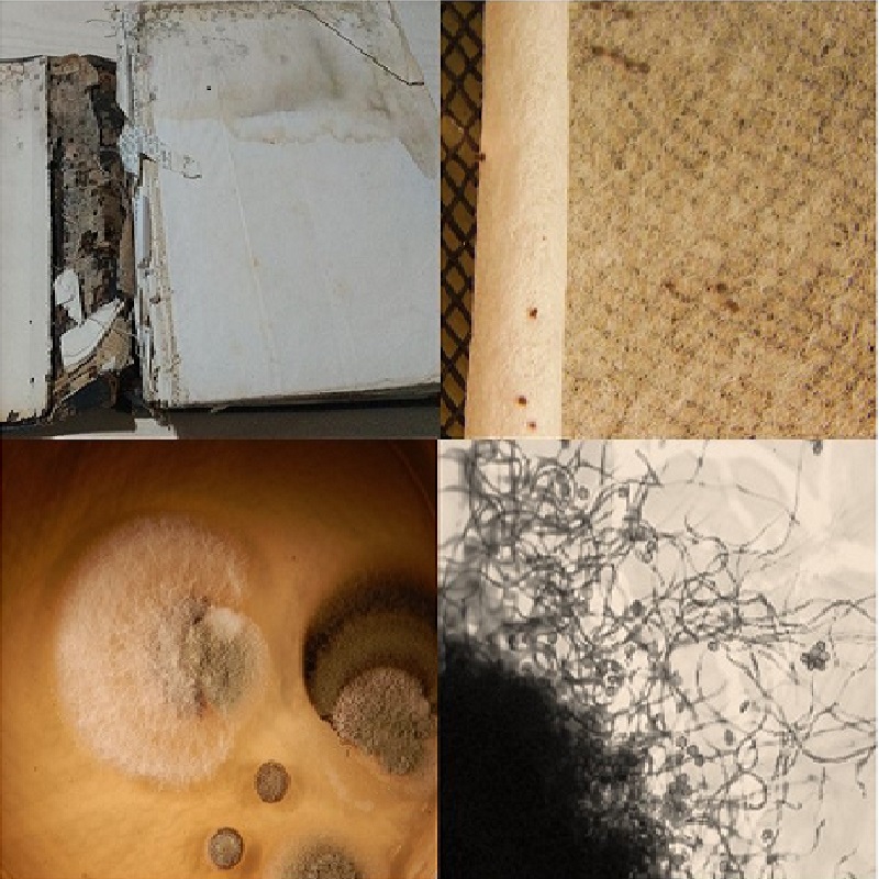
Pigmentos sintetizados por hongos negros y su impacto en el deterioro del patrimonio documental en papel
DOI:
https://doi.org/10.31055/1851.2372.v57.n2.36580Palabras clave:
biodeterioro, conservación, melaninas, hongos negros, papelResumen
Introducción y objetivos: Los documentos en papel custodiados en museos y bibliotecas pueden mostrar signos notorios de deterioro causados por la actividad de diferentes hongos. Algunos de los principales colorantes de origen fúngico que deterioran estéticamente este sustrato y afectan al patrimonio cultural en soporte celulósico son los pigmentos oscuros o melaninas. El objetivo del presente trabajo es brindar un panorama actualizado del estado de arte de los hongos negros que colonizan papel y las melaninas que sintetizan, ocasionando un daño importante al patrimonio documental, el cual es una fuente inigualable para la historia de los pueblos.
M&M: Se realizó una búsqueda bibliográfica de la información actualizada y disponible sobre los pigmentos oscuros que sintetizan diferentes hongos negros que deterioran papel. Para ello se analizaron 74 trabajos especializados en el tema; la mayoría de ellos de reciente publicación en revistas nacionales e internacionales.
Resultados: El conocimiento sobre la diversidad y las características de los pigmentos oscuros que son sintetizados por los hongos negros que deterioran papel es clave para desarrollar estrategias de prevención y remediación para eliminar estos pigmentos de soportes celulósicos con valor patrimonial. Este trabajo presenta información sobre los hongos negros que deterioran papel, los tipos de melaninas que pueden sintetizar, las estructuras donde se acumulan, y su contribución en el deterioro estético de los materiales mencionados
Conclusiones: Este conocimiento sirve de base para desarrollar nuevas estrategias de restauración que pudieran ser efectivas y sustentables y que aseguren la conservación preventiva de documentos históricos y obras de arte en papel.
Referencias
ALMENDROS, M. G., A. T. MARTINEZ, M. F. MARTÍNEZ & F. J. GONZÁLEZ-VILA. 1985. Degradative oxidation products of the melanin of Ulocladium atrum. Soil Biol. Biochem. 17: 723-726.
https://doi.org/10.1016/0038-0717(85)90052-5
ARAI, H. 2000. Foxing caused by Fungi: twenty-ve years of study. Inter. Biodet. Biodegradation 46: 181-188.
https://doi.org/10.1016/S0964-8305(00)00063-9
BABITSKAYA, V. G., V. V. SHCHERBA, T. V. FILIMONOVA, & E. A. GRIGORCHUK. 2000. Melanin pigments from the fungi Paecilomyces variotii and Aspergillus carbonarius. Appl. Biochem. Microbiol. 36: 128-133.
https://doi.org/10.1007/BF02737906
BÁRCENA, A., G. PETROSELLI, S. M. VELASQUEZ, J. M. ESTÉVEZ, R. ERRA-BALSELLS, P. A. BALATTI & M. C. N. SAPARRAT. 2015. Response of the fungus Pseudocercospora griseola f. mesoamericana to Tricyclazole. Mycol. Prog. 14:76.
https://doi.org/10.1007/s11557-015-1102-7
BÁRCENA, A., M. BRUNO, A. GENNARO, M. F. ROZAS, M. V. MIRÍFICO, P. A. BALATTI & M. C. N. SAPARRAT. 2018. Melanins from two selected isolates of Pseudocercospora griseola grown in-vitro: Chemical features and redox activity. J. Photochem. Photobiol. B. Biol. 186: 207-215.
https://doi.org/10.1016/j.jphotobiol.2018.07.019
BELL, A. A. & M. H. WHEELER. 1986. Biosynthesis and functions of fungal melanins. Annu. Rev. Phytopathol. 24: 411-451.
https://doi.org/10.1146/annurev.py.24.090186.002211
BELTRÁN-GARCÍA, M. J., F. M. PRADO, M. S. OLIVEIRA, D. ORTIZ-MENDOZA, A. C. SCALFO, A. PESSOA, M. H. G. MEDEIROS, J. F. WHITE & P. DI MASCIO. 2014. Singlet molecular oxygen generation by light-activated DHN-melanin of the fungal pathogen Mycosphaerella fijiensis in black Sigatoka disease of bananas. PLoS ONE 9:e91616.
https://doi.org/10.1371/journal.pone.0091616
BHARDWAJ, N. & I. K. BHATNAGAR. 2002. Microbial deterioration of paper paintings. Biodeterioration of Materials 2: 132-135.
BODDY, L. & J. HISCOX. 2016. Fungal ecology: principles and mechanisms of colonization and competition by saprotrophic fungi. Microbiology Spectrum 4: FUNK-0019-2016.
https://doi.org/10.1128/microbiolspec.FUNK-0019-2016
BORREGO ALONSO, S. F., & O. HERRERA BARRIOS.2021. Calidad micológica ambiental en archivos cubanos y su impacto en la salud del personal. An. Acad. Cienc. Cuba 11.
CALVO, A. M., R. A. WILSON, J. W. BOK & N. P. KELLER. 2002. Relationship between secondary metabolism and fungal development. Microbiol. Mol. Biol. Rev 66: 447-459.
https://doi.org/10.1128/MMBR.66.3.447-459.2002
CALVO, A. M. C., A. DOCTERS, M. V. MIRANDA & M. C. N. SAPARRAT. 2017. The use of gamma radiation for the treatment of cultural heritage in the Argentine National Atomic Energy Commission: past, present, and future. Top Curr Chem 375: 227-248.
https://doi.org/10.1007/s41061-016-0087-2
CAMACHO, E., R. VIJ, C. CHRISSIAN, R. PRADOS-ROSALES, D. GIL, R. N. O'MEALLY, R. J. B. CORDERO, R. N. COLE, J. M. McCAFFERY, R. E. STARK & A. CASADEVALL. 2019. The structural unit of melanin in the cell wall of the fungal pathogen Cryptococcus neoformans. J. Biol. Chem 294: 10471-10489.
https://doi.org/10.1074/jbc.RA119.008684
CHIEWCHANVIT, S., S. CHONGKAE, P. MAHANUPAB, J. D. NOSANCHUK, S. PORNSUWAN, N. VANITTANAKOM & S. YOUNGCHIM. 2017. Melanization of Fusarium keratoplasticum (F. solani Species Complex) during disseminated fusariosis in a patient with acute leukemia. Mycopathologia 182: 879-885.
https://doi.org/10.1007/s11046-017-0156-2
CHRISSIAN, C., E. CAMACHO, M. S. FU, R. PRADOS-ROSALES, S. CHATTERJEE, R. J. B. CORDERO, J. K. LODGE, A. CASADEVALL & R. E. STARK. 2020. Melanin deposition in two Cryptococcus species depends on cell-wall composition and flexibility. J. Biol. Chem 295: 1815-1828.
https://doi.org/10.1074/jbc.RA119.011949
DADACHOVA, E., R. A. BRYAN, X. HUANG, T. MOADEL, A. D. SCHWEITZER, P. AISEN, J. D. NOSANCHUK & A. CASADEVALL. 2007. Ionizing radiation changes the electronic properties of melanin and enhances the growth of melanized fungi. PLoS ONE 5: e457.
https://doi.org/10.1371/journal.pone.0000457
EVELEIGH, D. E. 1970. Fungal disfigurement of paper, and soft rot of cedar shingles. Applied Microbiology 19: 872-874.
https://doi.org/10.1128/am.19.5.872-874.1970
ELLIS, M. B. 1971. Dematiaceous Hyphomycetes. Commonwealth Mycological Institute. Kew, London.
ELLIS, M. B. 1976. More Dematiaceous Hyphomycetes. Commonwealth Mycological Institute, Kew, London.
FERNANDES, C., R. PRADOS-ROSALES, B. M. SILVA, A. NAKOUZI-NARANJO, M., ZUZARTE, S. CHATTERJEE, R. E. STARK, A. CASADEVALL & T. GONÇALVES. 2015. Activation of melanin synthesis in Alternaria infectoria by antifungal drugs. Antimicrob. Agents Chemother. 60 :1646-55.
https://doi.org/10.1128/AAC.02190-15
FERRÁNDIZ-PULIDO, C., M. T. MARTIN-GOMEZ, T. REPISO, C. JUÁREZ-DOBJANSCHI, B. FERRER, I. LÓPEZ-LERMA, G. APARICIO, C. GONZÁLEZ-CRUZ, F. MORESO, A. ROMAN & V. GARCÍA-PATOS. 2019. Cutaneous infections by dematiaceous opportunistic fungi: Diagnosis and management in 11 solid organ transplant recipients. Mycoses 62: 121-127.
https://doi.org/10.1111/myc.12853
FRANCO, E., M. I. TRONCOZO, M. BAEZ, M. V. MIRÍFICO, G. L. ROBLEDO, P. A. BALATTI & M. C. N. SAPARRAT. 2018. Fusarium equiseti LPSC 1166 and its in vitro role in the decay of Heterostachys ritteriana leaf litter. Folia Microbiologica 63: 169-179.
https://doi.org/10.1007/s12223-017-0541-8
FRANDSEN, R. J. N., S. A. RASMUSSEN, P. B. KNUDSEN, S. UHLIG, D. PETERSEN, E. LYSØE, C. H. H, H. GIESE & T. O. LARSEN. 2016. Black perithecial pigmentation in Fusarium species is due to the accumulation of 5-deoxybostrycoidin-based melanin. Scientific Reports 6: 26206.
https://doi.org/10.1038/srep26206
GESSLER, N. N., A. C. EGOROVA & T. A. BELOZERSKAYA. 2014. Melanin pigments of fungi under extreme environmental conditions. Appl. Biochem. Microbiol. 50: 105-113.
https://doi.org/10.1134/S0003683814020094
GMOSER, R, J. A. FERREIRA, P. R. LENNARTSSON & M. J. TAHERZADEH. 2017. Filamentous ascomycetes fungi as a source of natural pigments. Fungal Biol. Biotechnol. 4:4.
https://doi.org/10.1186/s40694-017-0033-2
HU, Y., X. HAO, J. LOU, P. ZHANG, J. PAN & X. ZHU. 2012. A PKS gene, pks-1, is involved in chaetoglobosin biosynthesis, pigmentation and sporulation in Chaetomium globosum. Sci. China Life Sci. 55: 1100-1108.
https://doi.org/10.1007/s11427-012-4409-5
KARAKASIDOU, K., K NIKOLOULI., G. D. AMOUTZIAS, A. PORNOU, CH. MANASSIS, G. TSIAMIS & D. MOSSIALOS. 2017. Microbial diversity in biodeteriorated Greek historical documents dating back to the 19th and 20th century: a case study. Microbiology Open 1: 11.
https://doi.org/10.1002/mbo3.596
KRAKOVÁ, L., K. CHOVANOVÁ, S. A. SELIM, A. ŠIMONOVIČOVÁ, A. PUŠKAROVÁ, A.D. MAKOVÁ & A. PANGALLO. 2012. Multiphasic approach for investigation of the microbial diversity and its biodegradative abilities in historical paper and parchment documents. Inter. Biodet. Biodegradation 70: 117-125.
https://doi.org/10.1016/j.ibiod.2012.01.011
LACEY, M. E. & J. S. WEST. 2006. The Air Spore. Springer. Dordrecht. https://doi.org/10.1007/978-0-387-30253-9
LECOINTE, K., M. CORNU, J. LEROY, P. COULON & B. SENDID. 2019. Polysaccharides cell wall architecture of Mucorales. Front. Microbiol. 10: 469.
https://doi.org/10.3389/fmicb.2019.00469
LEE D, E-H. JANG, M. LEE, S-W. KIM, Y. LEE, K-T. LEE & Y-S. BAHN. 2019. Unraveling melanin biosynthesis and signaling networks in Cryptococcus neoformans.
https://doi.org/10.1128/mBio.02267-19
LLORENTE, C, A. BÁRCENA, J. VERA BAHIMA, M. C. N. SAPARRAT, A. M. ARAMBARRI, M., F. ROZAS, M. V. MIRÍFICO & P. A. BALATTI. 2012. Cladosporium cladosporioides LPSC 1088 produces the 1,8-dihydroxynaphthalene-melanin-like compound and carries a putative pks gene. Mycopathologia 174: 397-408.
https://doi.org/10.1007/s11046-012-9558-3
MALLO, A. C., D. S. NITIU, L. A. ELÍADES & M. C. N. SAPARRAT. 2017. Fungal degradation of cellulosic materials used as support for cultural heritage. Int. J. Conserv. Sci. 8: 619-632.
MEDINA, R., C. G. LUCENTINI, M. E. E. FRANCO, G. PETROSELLI, J. A. ROSSO, R. ERRA-BALSELLS, P. A. BALATTI & M. C. N. SAPARRAT. 2018. Identification of an intermediate for 1,8- dihydroxynaphthalene-melanin synthesis in a race-2 isolate of Fulvia fulva (syn. Cladosporium fulvum). Heliyon.
https://doi.org/10.1016/j.heliyon.2018.e01036
MELO, D., S. O. SEQUEIRA, J. A. LOPES & M. F. MACEDO. 2019. Stains versus colourants produced by fungi colonising paper cultural heritage: A review. J. Cult. Herit 35: 161-182. https://doi.org/10.1016/j.culher.2018.05.013
MELO, D. C. 2017. Fungal Stains on Paper: Melanins produced by fungi. Thesis for the Master degree in Conservation and Restoration. Monte de Caparica, Lisboa: Departamento de Conservação e Restauro, Mestrado em Conservação e Restauro, Faculdade de Ciências e Tecnologia, Universidade Nova de Lisboa. 57 p.
MESQUITA, N., A. PORTUGAL, S. VIDEIRA, S. RODRÍGUEZ-ECHEVERRÍA, A. M. L. BANDEIRA, M. J. A. SANTOS & H. FREITAS. 2009. Fungal diversity in ancient documents. A case study on the Archive of the University of Coimbra. Int. Biodeterior. Biodegradation. 63: 626-629.
https://doi.org/10.1016/j.ibiod.2009.03.010
MICHAELSEN, A., F. PINZARI, K. RIPKA, W. LUBITZ & G. PIÑAR. 2006. Application of molecular techniques for identification of fungal communities colonizing paper material. Inter. Biodet. Biodegradation 58:133-141.
https://doi.org/10.1016/j.ibiod.2006.06.019
MICHAELSEN, A., G. PIÑAR & F. PINZARI. 2010. Molecular and microscopical investigation of the microflora inhabiting a deteriorated Italian manuscript dated from the thirteenth century. Microbial Ecology 60: 69-80.
https://doi.org/10.1007/s00248-010-9667-9
NOL, L., Y. HENIS & R. G. KENNETH. 2001. Biological factors of foxing in postage stamp paper. Inter. Biodet. Biodegradation 48: 98-104.
https://doi.org/10.1016/S0964-8305(01)00072-5
NG, K.P., S. M. YEW, CH. L. CHAN, T. S. SOO-HOO, S. L. NA, H. HASSAN, Y. F. NGEOW, CH. CH. HOH, K. W. LEE & W. Y. YEE. 2012. Sequencing of Cladosporium sphaerospermum, a dematiaceous fungus isolated from blood culture. Eukaryotic Cell 11: 705-706.
https://doi.org/10.1128/EC.00081-12
NITIU, D. S., A. C. MALLO & M. C. N. SAPARRAT. 2020. Fungal melanins that deteriorate paper cultural heritage: an overview. Mycologia. 112: 859-870. https://doi.org/10.1080/00275514.2020.1788846
NOSANCHUK, J. D., R. E. STARK & A. CASADEVALL. 2015. Fungal melanin: what do we know about structure? Front. Microbiol. 6: 1463.
https://doi.org/10.3389/fmicb.2015.01463
PAL, A. K., D. H. GAJJAR & A. R. VASAVADA. 2014. DOPA and DHN pathway orchestrate melanin synthesis in Aspergillus species. Medical Mycology 52: 10-18.
https://doi.org/10.3109/13693786.2013.826879
PALONEN, E. K., S. RAINA, A. BRANDT, J. MERILUOTO, T. KESHAVARZ & J. T. SOINI. 2017. Melanisation of Aspergillus terreus-Is butyrolactone I involved in the regulation of both DOPA and DHN types of pigments in submerged culture? Microorganisms 5: 22.
https://doi.org/10.3390/microorganisms5020022
PANGALLO, D., K. CHOVANOVA, A. ŠIMONOVIČOVÁ & P. FERIANIC. 2009. Investigation of microbial community isolated from indoor artworks and air environment: identification, biodegradative abilities, and DNA typing. Can. J. Microbiol. 55: 277-287.
https://doi.org/10.1139/w08-136
PAVON FLORES, S. C. 1976. Gamma radiation as fungicide and its effects on paper. Bulletin of the American Institute for Conservation of Historic and Artistic Works 16: 15-44.
https://doi.org/10.1179/019713676806029384
PEREZ-CUESTA, U., L. APARICIO-FERNANDEZ, X. GURUCEAGA, L. MARTIN-SOUTO, A. ABAD-DIAZ-DE-CERIO, A. ANTORAN, I. BULDAIN, F. L. HERNANDO, A. RAMIREZ-GARCIA & A. REMENTERIA. 2019. Melanin and pyomelanin in Aspergillus fumigatus: from its genetics to host interaction. Int. Microbiol. https://doi.org/10.1007/s10123-019-00078-0
PICCOLO, A. 1996. Humic substances in terrestrial ecosystems. 1st ed. Elsevier, Amsterdam,
PINHEIRO, A. C, M. F. MACEDO, V. JURADO, C. SAINZ JIMENEZ, C. VIEGAS, J. BRANDÃO & L. ROSADO. 2011. Mould and yeast identification in archival settings: Preliminary results on the use of traditional methods and molecular biology options in Portuguese archives. Int. Biodeterior. Biodegradation. 65: 619-627.
https://doi.org/10.1016/j.ibiod.2011.02.008
PINZARI, F. & M. MONTANARI. 2011. Mould growth on library materials stored compactus-type shelving units. In: ABDUL-WAHAB SA, ed. Sick Building Syndrome in Public Buildings and Workplaces, p. 196-203. Springer, Berlin.
https://doi.org/10.1007/978-3-642-17919-8_11
POMBEIRO-SPONCHIADO, S. R., G. S. SOUSA, J. C. ANDRADE, H. F. LISBOA & R. C. GONÇALVES. 2017. Production of melanin pigment by fungi and its biotechnological applications. In BLUMENBERG, M. (ed.), Melanin, p; 45-75. InTechOpen, London.
QUAN, Y., B. G. VAN DEN ENDE, D. SHI, F. X. PRENAFETA-BOLDÚ, Z. LIU, A. M. S. AL-HATMI, S. A. AHMED, P. E. VERWEIJ, Y. KANG & S. DE HOOG. 2019. A comparison of isolation methods for black fungi degrading aromatic toxins. Mycopathologia 184: 653-660.
https://doi.org/10.1007/s11046-019-00382-3
RAKOTONIRAINY, M. S., E HEUDE & B. LAVÉDRINE. 2007. Isolation and attempts of biomolecular characterization of fungal strains associated to foxing on a 19th century book. Journal of Cultural Heritage 8 : 126-133.
https://doi.org/10.1016/j.culher.2007.01.003
RAO, M. P. N., M. XIAO & W. J. LI. 2017. Fungal and bacterial pigments: Secondary metabolites with wide application. Front. Microbiol. 8 :1113.
https://doi.org/10.3389/fmicb.2017.01113
REIS-MENESES, A. A., W. GAMBALE, M. C. GIUDICE & M. A. SHIRAKAWA. 2011. Accelerated testing of mold growth on traditional and recycled book paper. Int. Biodeterior. Biodegradation 65: 423-428. https://doi.org/10.1016/j.ibiod.2011.01.006
ROJAS, T. I., M. J. AIRA, A. BATISTA, I. L. CRUZ & S. GONZÁLEZ. 2012. Fungal biodeterioration in historic buildings of Havana (Cuba). Grana 51 (1): 44-51.
https://doi.org/10.1080/00173134.2011.643920
RUIBAL, C., G. PLATAS & G. F. BILLS. 2008. High diversity and morphological convergence among melanised fungi from rock formations in the Central Mountain System of Spain. Persoonia 21: 93-110.
https://doi.org/10.3767/003158508X371379
RUISI, S., D. BARRECA, L. SELBMANN, L. ZUCCONI & S. ONOFRI. 2007. Fungi in Antarctica. Rev. Environ. Sci. Biotechnol. 6: 127-141.
https://doi.org/10.1007/s11157-006-9107-y
SAPARRAT, M. C. N., G. FERMOSELLE, S. STENGLEIN, M. AULICINO & P. A. BALATTI. 2009. Pseudocercospora griseola causing angular leaf spot on Phaseolus vulgaris produces 1,8-dihydroxynaphthalene-melanin. Mycopathologia 168: 41-47.
https://doi.org/10.1007/s11046-009-9194-8
SARI, E., L. ISERI, M. KOÇAK & D. YILDIZ. 2015. Is it Subungual Melanoma? Fungal melanonychia due to Phoma glomerata. Cukurova Medical Journal 40: 162-165.
https://doi.org/10.17826/cutf.44511
SCHMALER-RIPCKE, J., V. SUGAREVA, P. GEBHARDT, R. WINKLER, O. KNIEMEYER, T. HEINEKAMP & A. A. BRAKHAG. 2009. Production of pyomelanin, a second type of melanin, via the tyrosine degradation pathway in Aspergillus fumigatus. Appl. Environ. Microbiol. 75: 493-503.
https://doi.org/10.1128/AEM.02077-08
SMITH, D. F. Q. & A. CASADEVALL. 2019. The role of melanin in fungal pathogenesis for animal hosts. In: RODRIGUES M. (ed.), Fungal Physiology and Immunopathogenesis. Curr. Top. Microbiol. Immunol. Vol. 422, pp. 1-30. Springer, Cham, Switzerland.
https://doi.org/10.1007/82_2019_173
STALEY, J. T., F. E. PALMER & J. B. ADAMS. 1982. Microcolonial fungi: Common inhabitants on desert rocks? Science 215: 1093-1095. https://doi.org/10.1126/science.215.4536.1093
STERFLIGER, K. 2010. Fungi: Their role in deterioration of cultural heritage. Fungal Biology Reviews 24: 47-55.
https://doi.org/10.1016/j.fbr.2010.03.003
SUN, S., X. ZHANG, S. SUN, L. ZHANG, S. SHAN & H. ZHU. 2016. Production of natural melanin by Auricularia auricula and study on its molecular structure. Food Chemistry 190:801-807.
https://doi.org/10.1016/j.foodchem.2015.06.042
SZCZEPANOWSKA, H. & A. R. CAVALIERE. 2012. Conserving our cultural heritage: The role of fungi in biodeterioration. In: JOHANNING E, MOREY P, AUGER P. (eds.), Bioaerosols Fungi, Bacteria, Mycotoxins in Indoor and Outdoor Environments and Human Health, pp. 293-309. Fungal Research Group, Albany
TOLEDO, A. V, M. E. E. FRANCO, S. M. Y. LÓPEZ, M. I. TRONCOZO, M. C. N. SAPARRAT & P. A. BALATTI. 2017. Melanins in fungi: Types, localization and putative biological roles. Physiol. Mol. Plant Pathol. 99: 2-6. https://doi.org/10.1016/j.pmpp.2017.04.004
TUDOR, D., S. C. ROBINSON & P. A. COOPER. 2012. The influence of moisture content variation on fungal pigment formation in spalted wood. AMB Express 2:69.
https://doi.org/10.1186/2191-0855-2-69
TUDOR, D. 2013. Fungal pigment formation in wood substrate. Ph.D. Thesis, University of Toronto, Toronto, ON, Canada, 2013.
WAINWRIGHT, M., T. A. ALI & F. BARAKAH. 1993. A review of the role of oligotrophic micro-organisms in biodeterioration. Int. Biodet. Biodegradation 31: 1-13.
https://doi.org/10.1016/0964-8305(93)90010-Y
WOUDENBERG, J. H. C., M. MEIJER, J. HOUBRAKEN, & R. A. SAMSON. 2017. Scopulariopsis and scopulariopsis-like species from indoor environments. Studies in Mycology 1-35.
https://doi.org/10.1016/j.simyco.2017.03.001
WALKER, C. A., B. L. GÓMEZ, H. M. MORA-MONTES, K. S. MACKENZIE, C. A. MUNRO, A. J. BROWN, N. A. GOW, C. C. KIBBLER & F. C. ODDS. 2010. Melanin externalization in Candida albicans depends on cell wall chitin structures. Eukaryotic Cell 9: 1329-1342.
https://doi.org/10.1128/EC.00051-10
ZHENG, W., B. S. CAMPBELL, B. M. MCDOUGALL & R. J. SEVIOUR. 2008. Effects of melanin on the accumulation of exopolysaccharides by Aureobasidium pullulans grown on nitrate. Bioresource Technology 16: 7480-6.
https://doi.org/10.1016/j.biortech.2008.02.016
ZOTTI, M, A. FERRONI & P. CALVINI. 2008. Microfungal biodeterioration of historic paper: Preliminary FTIR and microbiological analyses. Int. Biodet. Biodegradation. 62: 186-194.

Publicado
Número
Sección
Licencia
Derechos de autor 2022 Daniela Silvana Nitiu, Andrea Cecilia Mallo, Mario Carlos Nazareno Saparrat

Esta obra está bajo una licencia internacional Creative Commons Atribución-NoComercial-SinDerivadas 4.0.
El Bol. Soc. Argent. Bot.:
- Provee ACCESO ABIERTO y gratuito inmediato a su contenido bajo el principio de que hacer disponible gratuitamente la investigación al público, lo cual fomenta un mayor intercambio de conocimiento global.

- Permite a los autores mantener sus derechos de autor sin restricciones.
- El material publicado en Bol. Soc. Argent. Bot. se distribuye bajo una licencia de Creative Commons Atribución-NoComercial-CompartirIgual 4.0 Internacional.


