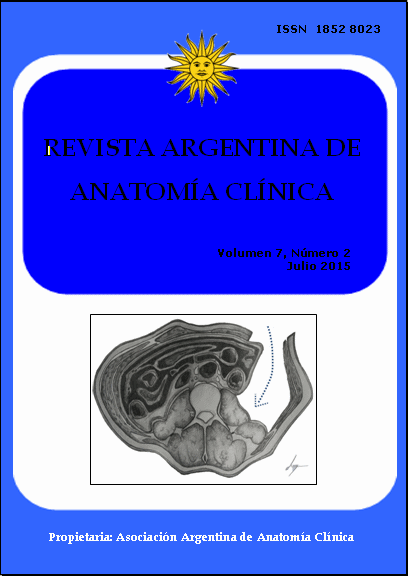
DRENAJE QUIRÚRGICO EXTRAPERITONEAL DE ABSCESO DEL PSOAS: FUNDAMENTO ANATÓMICO. Drenaje quirúrgico extraperitoneal de absceso del psoas: Fundamento anatómico
DOI:
https://doi.org/10.31051/1852.8023.v7.n2.14174Keywords:
retroinguinal space, psoas muscle, iliac muscle, retroperitoneal region, clinical anatomy, espacio retroinguinal, músculo psoas, músculo ilíaco, espacio extraperitoneal, anatomía clínicaAbstract
El espacio extraperitoneal se encuentra delimitado por el peritoneo parietal y las paredes de la cavidad abdómino-pélvica. Al igual que la cavidad peritoneal este espacio puede ser asiento de diversas colecciones, como ser hematomas, tumores y supuración. Con el advenimiento de las nuevas técnicas de imagen, se ha contribuido no solo al mejor diagnóstico de estas patologías sino también a su mejor manejo. El objetivo de este trabajo es mostrar la anatomía del abordaje extraperitoneal del comparti-miento del psoas y su aplicación al tratamiento de un paciente. Para esto se utilizaron 5 cadáveres adultos fijados previamente en solución en base a formol. Se realizó disección bilateral de la pared antero-lateral del abdomen reclinando la bolsa peritoneal para a continuación abordar el compartimiento del músculo psoas. Este conocimiento fue utilizado en el tratamiento quirúrgico de una paciente que consultó por un absceso del compartimiento del psoas derecho. En las preparaciones cadavéricas, se observó cómo al rebatir el peritoneo parietal se expone la totalidad del compartimiento muscular del psoas. Este procedi-miento fue realizado a la paciente consiguiendo el drenaje completo de la cavidad abscedada, quien tuvo una buena evolución y fue dada de alta a los 7 días. Los hallazgos demuestran una vez más como el conocimiento anatómico sigue estando vigente en la práctica clínica, siendo la comprensión del espacio extraperitoneal fundamental no solo para el anatomista sino también para el cirujano.
The retroperitoneal space is bounded by the parietal peritoneum and the posterior abdominal wall. Just like the peritoneal cavity, this region can host multiple effusions such as hematomas, tumors and suppuration. With the development of new radiological technics, both diagnosis and management of these conditions has improved. The purpose of this paper is to demonstrate the anatomy of the extraperitoneal approach of the psoas compartment and its application to a patient´s surgical treatment. For this purpose 5 formalin-fixed adult cadavers were used. Bilateral dissection of the antero-lateral abdominal wall was performed in every specimen. Once the parietal peritoneum was mobilized the psoas compartment was approached. This knowledge was used during the surgical treatment of a patient who attended to the emergency room with a right psoas compartment abscess. In the cadaveric specimens, the psoas muscular compartment was approached after mobilizing the parietal peritoneum medially. This procedure was carried out in the patient resulting in complete drainage of the purulent effusion. The patient had complete relief of the symptoms and was discharged 7 days after the procedure. These findings show that the anatomic knowledge is still important in clinical practice. Understanding the extraperitoneal space is crucial for both anatomists and surgeons.
References
Afaq A, Jain BK, Dargan P, Bhattacharya SK, Rauniyar RK, Kukreti R. 2002. Surgical drainage of primary iliopsoas abscess-safe and cost-effective treatment. Trop Doct, 32: 133–35.
Bendavid R. 2001. Abdominal Wall Hernias. Nueva York: Springer Science+Business Media, pág: 101–06.
Brick WG, Colborn GL, Gadacz TR, Skandalakis JE. 1995. Crucial anatomic lessons for laparoscopic herniorrhaphy. Am Surg, 61: 172–77.
Cantasdemir M, Kara B, Cebi D, Selcuk ND, Numan F. 2003. Computed tomography-guided percutaneous catheter drainage of primary and secondary iliopsoas abscesses. Clin Radiol, 58: 811–15.
Colborn GL, Brick WG, Gadacz TR, Skandalakis JE. 1995. Inguinal anatomy for laparoscopic herniorrhaphy, Part I: The normal anatomy. Surg Rounds, 18: 189–98.
Colborn GL, Skandalakis JE. 1998. Laparoscopic inguinal anatomy. Hernia, 2: 179–91.
Couinaud C. 1963. Anatomie de l´abdomen. Paris: G. Doin et Cie, Edit, pág: 1–847.
De la Torre González D, Góngora López J, Pérez Meave JA, Guerrero Beltrán L, Miranda Gómez D, Bon Villareal JR. 2006. Mal de Pott. Diagnóstico y tratamiento del paciente. Rev Hosp Jua Mex, 3: 96–100.
Desandre AR, Cottone FJ. 1995. Iliopsoas abscess: etiology, diagnosis, and treatment. Am Surg, 61: 1087–91.
Gruenwald I, Abrahamson J, Cohen O. 1992. Psoas abscess: case report and review of the literature. J Urol, 147: 1624–26.
Hureau J, Agossou-Voyeme AK, Germain M, Pradel J. 1991. Les espaces interpariéto-peritonéaux postérieurs ou les espaces rétropéritonéaux: anatomie topographique normal. J Radiol, 72: 101–16.
Hureau J, Pradel J, Agossou-Voyeme AK, Germain M. 1991. Les espaces interpariéto-péritonéaux postérieurs ou les espaces rétropéritonéaux: anatomie tomodensito-métrique pathologique. J Radiol, 72: 205–27.
Lai YC, Lin PC, Wang WS, Lai JI. 2011. An Update on Psoas Abscess: An 8-Year Experience and Review of the Literature. International Journal of Gerontology, 5: 75–79.
Matin SF, Gill IS. 2002. Laparoscopic radical nephrectomy: retroperitoneal versus trans-peritoneal approach. Curr Urol Rep, 3: 164–71.
Meyer HI. 1934. The reaction of retroperitoneal tissue to infection. Ann Surg, 99: 246–50.
Meyers MA, Charnsangavej C, Oliphant M. 2011. Meyer´s Dynamic Radiology of the Abdomen. Nueva York: Springer-Verlag, pag: 1–800.
Mynter H. 1881. Acute psoitis. Buffalo Med Surg J, 21: 202–10.
Navarro López V, Ramos JM, Mesenguer V, Pérez Arellano JL, Serrano R, García Ordoñez MA, Peralta G, Boix V, Pardo J, Conde A, Salgado F, Gutiérrez F. 2009. Microbiology and outcome of iliopsoas abscess in 124 patients. Medicine (Baltimore), 88: 120–30.
Paturet G. 1951. Traité d´Anatomie Humaine. Vol 1. Paris: Masson et Cie Edit, pag: 1–758.
Pérez-Fernández S, de la Fuente-Aguado J, Fernández-Fernández FJ, Rubianes-González M, Pérez-Arguelles BS, Martínez-Vázquez C. 2006. Abscesos del psoas. Una perspectiva actual. Enferm Infecc Microbiol Clin, 24: 313–18.
Pró E. 2012. Anatomía Clínica. Buenos Aires: Médica Panamericana, pág: 1–963.
Ricci MA, Rose FB, Meyer KK. 1986. Pyogenic psoas abscess: worldwide variations in etiology. World J Surg, 10: 834–43.
Rouviere H, Delmas A. 1999. Anatomía Humana, Tomo Segundo. Barcelona: Editorial Masson, pag: 369–74.
Santaella RO, Fishman EK, Lipsett PA. 1995. Primary vs. secondary iliopsoas abscess. Arch Surg, 130: 1309–13.
Skandalakis JE, Colborn GL, Weidman TA, Foster RS, Kingsnorth AN, Skandalakis LJ, Skandalakis PN, Mirilas PS. 2004. Skandalakis´ Surgical Anatomy: The Embryologic and Anatomic Basis of Modern Surgery. Grecia: Paschalidis Medical Publications, pag: 1–986.
Stevenson EO, Ozeran RS. 1969. Retro-peritoneal space abscesses. Surg Gynecol Obstet, 128: 1202–08.
Testut L, Jacob O. 1979. Tratado de Anatomía topográfica, Tomo II. Barcelona: Editorial Salvat, pag: 332–38.
Thornton FJ, Kandiah SS, Monkhouse WS. 2001. Helical CT evaluation of the perirenal space and its boundaries: a cadaveric study. Radiology, 218: 659–63.
Tosenovsky P, Janousek L, Lipar K, Moravec M. 2003. Left retroperitoneal versus trans-peritoneal approach for abdominal aortic surgery – retrospective comparison of intra-operative and postoperative data. Bratisl Lek Listy, 104: 325–55.
Yacoud WN, Sohn HJ, Chan S, Petrosvan M, Vermaire HM, Kelso RL, Towfigh S, Mason RJ. 2008. Psoas abscess rarely requires surgical intervention. Am J Surg, 196: 223–27.
Downloads
Published
Issue
Section
License
Authors retain copyright and grant the journal right of first publication with the work simultaneously licensed under a Creative Commons Attribution License that allows others to share the work with an acknowledgement of the work's authorship and initial publication in this journal. Use restricted to non commercial purposes.
Once the manuscript has been accepted for publications, authors will sign a Copyright Transfer Agreement to let the “Asociación Argentina de Anatomía Clínica” (Argentine Association of Clinical Anatomy) to edit, publish and disseminate the contribution.


